Parkinson’s Disease & Multiple System Atrophy Imaging for Clinical Trials
Regional brain volumes, cortical thickness, DAT SPECT, VMAT2 PET, FDG PET, and DTI for Parkinson's Disease and Multiple System Atrophy clinical trials.
Parkinson's Disease & MSA Imaging in Clinical Trials Overview
Multi-modality imaging is widely used in early and late phase clinical trials of Parkinson's Disease (PD) and Multiple System Atrophy (MSA). Key uses of imaging include:
- MRI
- Eligibility Reads
- Safety Reads
- Quantitative measures for efficacy endpoints (e.g. volumetric MRI, diffusion MRI, neuromelanin imaging)
- PET & SPECT
- Eligibility & Study Population Enrichment
- Quantitative measures for efficacy endpoints (e.g. SUVR, SBR)
The successful use of neuroimaging in clinical trials of PD and MSA requires the seamless combination of rigorous study operations, radiologic reads with robust data tracking, and state-of-the-art, validated image processing & analysis software to derive quantitative measures from high-quality images.
Imaging Biomarkers for Parkinson's Disease & Multiple System Atrophy
-
- DaTscan (DAT SPECT)
- VMAT2 PET
- FDG PET
- Volumetric MRI (regional volumes)
- Diffusion Imaging (e.g. DTI, NODDI, free water imaging)
- Neuromelanin Imaging
Highlights from Our Parkinson's Disease & Multiple System Atrophy Imaging Research
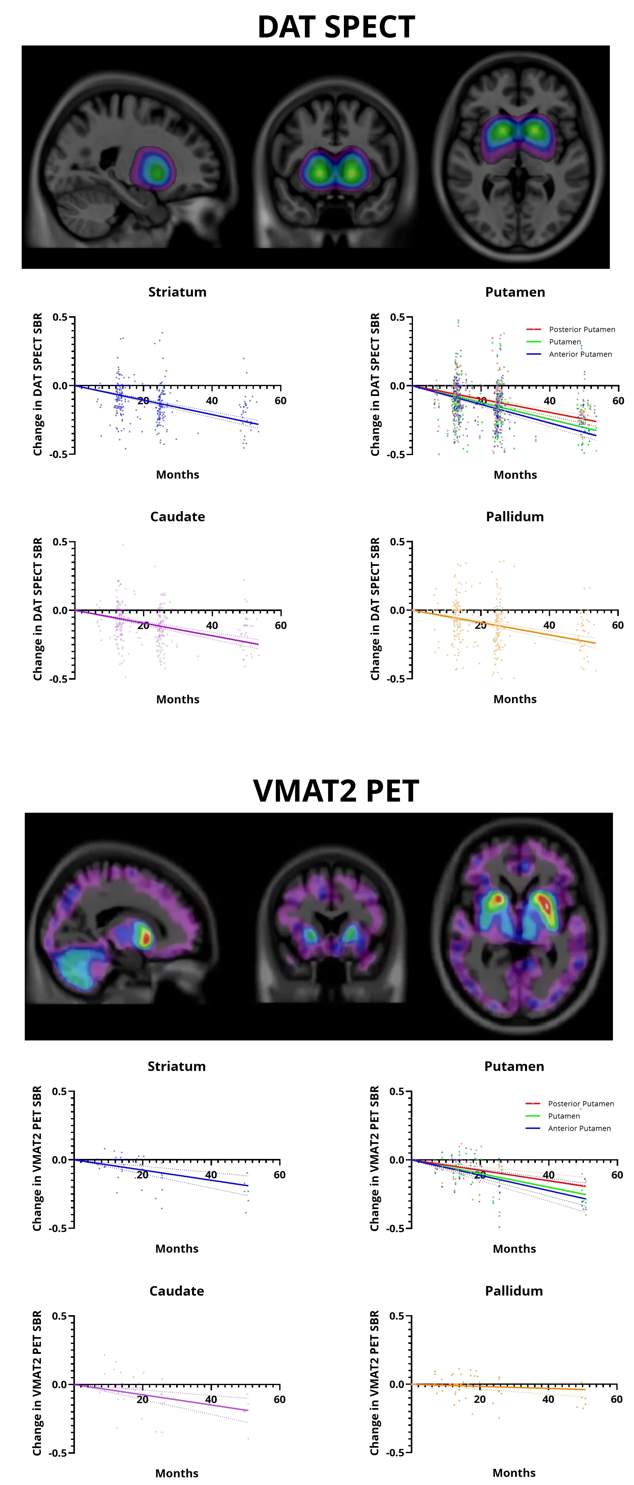
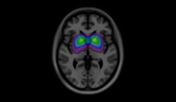
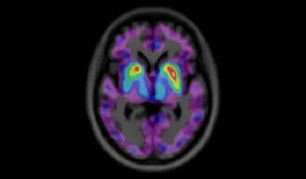
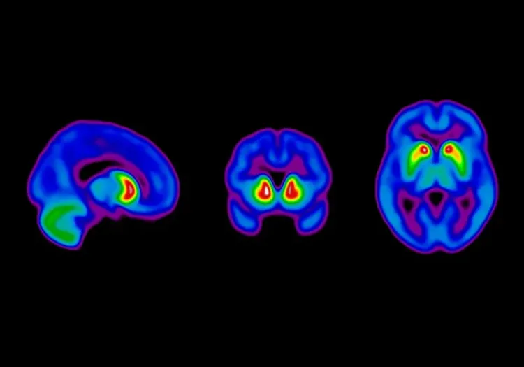
The thresholded DAT SPECT image, combined with 3D T1-weighted MRI, shows dopaminergic terminal density in PD patients at baseline. The gray matter masked VMAT2 PET image highlights higher spatial resolution than DAT SPECT. Scatter plots for both reveal a gradual decrease in SBR signals in the striatum, putamen, caudate, and pallidum over 48 months.
Dopaminergic Imaging in Parkinson's Disease
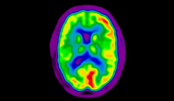
Neuroimaging biomarkers play a critical role in clinical trials for Parkinson's disease (PD) by providing objective, non-invasive tools to assess disease progression, evaluate treatment efficacy, and identify individuals at risk. Imaging modalities, such as MRI, PET, and SPECT, enable investigators to visualize structural, metabolic, and molecular brain changes, including dopamine system integrity and neurodegeneration. By offering measurable insights into key pathological processes, neuroimaging biomarkers enhance trial design, improve patient stratification, and facilitate earlier intervention strategies, ultimately advancing the development of targeted therapies for Parkinson's disease.
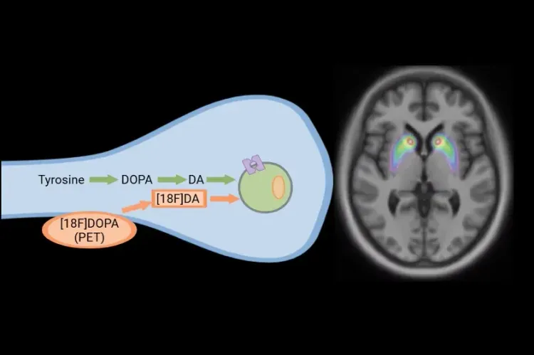
Dopaminergic neurodegeneration is a hallmark of PD, and imaging techniques, such as dopamine transporter single photon emission computed tomography (DAT SPECT) and vesicular monoamine transporter type 2 positron emission tomography (VMAT2 PET), are widely used to assess dopaminergic terminal loss. While DAT SPECT is well-established and broadly available, VMAT2 PET offers superior spatial resolution and enhanced ability to detect dopaminergic degeneration. Our analysis of the Parkinson's Progression Markers Initiative (PPMI) database compares the effectiveness of these two imaging modalities in tracking PD progression and determining optimal clinical trial sample sizes.
Our study employs our PIANO™ image processing platform to achieve high-throughput and precise registration of SPECT and PET scans to MRI, thereby enabling accurate region of interest analysis. This approach effectively shows longitudinal changes in dopaminergic terminal density within the putamen, highlighting its potential as a reliable marker for tracking disease progression.
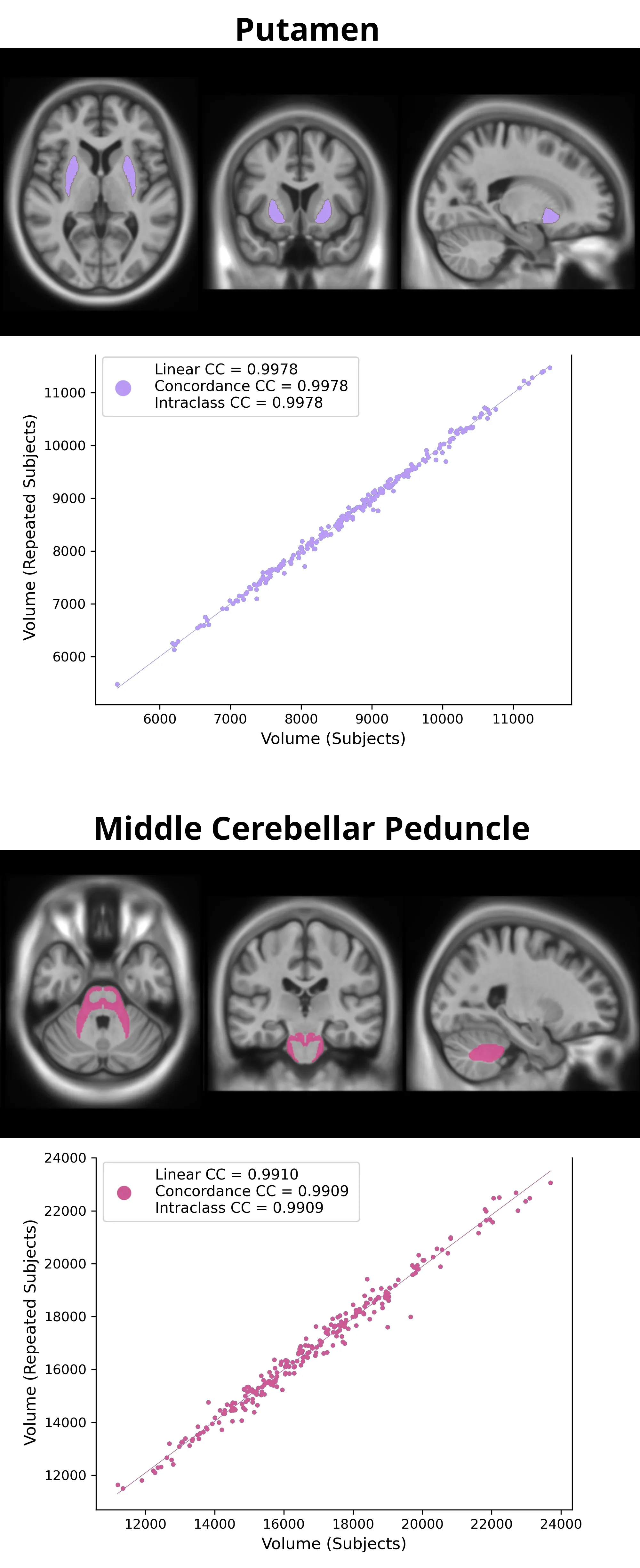
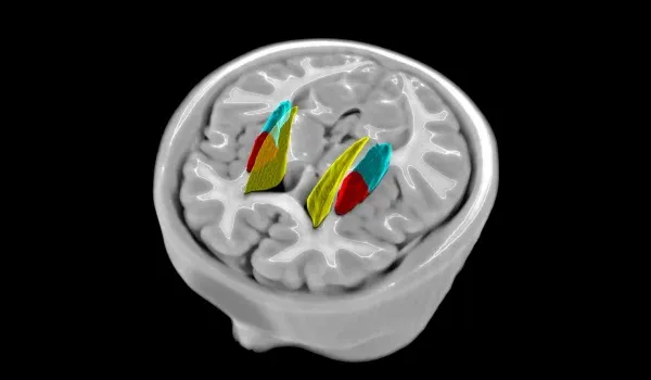
The anatomical MR images highlight the Putamen and Middle Cerebellar Peduncle regions-of-interest. The accompanying scatter plots display the regional volumes assessed across repeated scan sets. Each dot represents an individual subject, demonstrating the strong correlation between independently processed images. The observed correlation coefficient exceeds 0.99, underscoring the exceptionally high test-retest reliability of PIANO™ volumetric assessments.
Regional Volumetry in Multiple System Atrophy
Neuroimaging biomarkers are vital in clinical trials for Multiple System Atrophy (MSA), providing objective tools to assess disease progression, evaluate treatment efficacy, and identify at-risk individuals. MR imaging is widely used to visualize structural brain changes, offering valuable insights into MSA-related neurodegeneration. The current MSA diagnostic criteria require imaging to confirm the clinical diagnosis, underscoring the role of imaging biomarkers in MSA studies (Liu, 2024).
Brain atrophy is a prominent feature of MSA, with significant volume loss observed in the putamen, pons, and middle cerebellar peduncle (MCP). These regions are crucial for diagnosing MSA and monitoring progression. Notably, cerebellar atrophy is strongly linked to the MSA-C subtype, helping differentiate it from MSA-P, where the putamen is more severely affected (Gilman, 2008). MSA progresses rapidly, often advancing over 5 to 10 years. In patients with an average disease duration of four years, atrophy can be detected within just 12 months (Krismer, 2024). Identifying these changes early is vital for evaluating treatment effects in clinical trials.
Accurate and precise volumetric assessments are crucial for tracking subtle structural changes, but variability in image acquisition and processing can undermine reliability. Using data from the PPMI study, we have employed Biospective’s PIANO™ image processing platform, designed to minimize measurement variability and optimize volumetric assessment. Results demonstrate exceptionally low methodological variability, with correlation coefficients exceeding 0.99, enhancing our sensitivity to subtle volumetric changes and improving the ability to detect meaningful treatment effects. By reducing variability, our platform also optimizes sample size requirements, ultimately supporting more efficient clinical trial designs.
Learn more about the use of neuroimaging in clinical trials of Parkinson's Disease, Multiple System Atrophy, and our imaging biomarkers.
Discover more of our Therapeutic Areas
Related Content
Up-to-date information on Parkinson’s Disease and Multiple System Atrophy Imaging and best practices related to the evaluation of therapeutic agents in clinical trials.

MRI in Clinical Trials of Multiple System Atrophy (MSA)
This resource provides an overview of the utility of volumetric MRI and diffusion-weighted imaging (DWI) as biomarkers in research studies of MSA.
Dopaminergic Imaging in Parkinson's Disease
DAT SPECT and VMAT2 PET analysis in a natural history study of Parkinson's Disease.
[18F]DOPA PET in Parkinson’s Disease Clinical Trials
How [18F]DOPA PET is used to monitor disease progression & response to therapeutic intervention in Parkinson’s disease & movement disorders clinical trials.
Computational Method for Harmonization of Brain PET Images
A new algorithm for standardizing the spatial resolution of brain positron emission tomography (PET) images in multicenter clinical trials without phantoms.