Dopaminergic Imaging in Parkinson's Disease
Last Updated Date: March 13, 2025
Authors: Authors: Jean-Philippe Coutu, Ph.D., Simone P. Zehntner, Ph.D., Felix Carbonell, Ph.D., Alex P. Zijdenbos, Ph.D., Barry J. Bedell, M.D., Ph.D., for the Parkinson’s Progression Markers Initiative*
Key Takeaways
- DAT SPECT is a well-established and widely available clinical tool that provides valuable insights into dopamine transporter density. However, VMAT2 PET imaging offers superior spatial resolution and a more precise assessment of dopamine storage function.
- Despite not showing significant voxelwise effects after 12 months, VMAT2 PET regional SBR analysis detected significant effects after 12 months, as early as DAT SPECT, and in both cases effects were strongest in the putamen.
- With 12 to 24 months of follow-up, sample sizes estimated to be needed in a clinical trial are less than 50 participants per arm for DAT SPECT, and as few as 10 subjects per arm with VMAT2 PET.
- Both DAT SPECT and VMAT2 PET imaging modalities can reliably track PD progression and may serve as valuable biomarkers in clinical trials evaluating disease-modifying therapies.
Dopaminergic neurodegeneration is a hallmark of Parkinson's disease (PD), and imaging techniques are essential for assessing disease progression and evaluating therapeutic efficacy in clinical trials. Dopamine transporter single photon emission computed tomography (DAT SPECT) and vesicular monoamine transporter type 2 positron emission tomography (VMAT2 PET) are two primary imaging modalities used to visualize and quantify dopaminergic neuron loss. While DAT SPECT is widely available and clinically validated, VMAT2 PET offers superior spatial resolution and a more direct assessment of dopamine storage capacity. The comparative efficacy of these imaging techniques in tracking PD progression and determining optimal clinical trial sample sizes remains an area of investigation.
This study aimed to compare the performance of DAT SPECT and VMAT2 PET in tracking dopaminergic neurodegeneration in PD patients using data from the Parkinson's Progression Markers Initiative (PPMI). We also sought to determine the sample sizes required to detect significant therapeutic effects in PD clinical trials utilizing these imaging biomarkers.
Data were sourced from the PPMI, a multicenter longitudinal study designed to identify biomarkers for Parkinson's disease. DAT SPECT and VMAT2 PET imaging data were collected at baseline and follow-up intervals of 6, 12, 24, and 48 months. DAT SPECT was assessed in healthy controls (n=37) and PD patients (n=134), while VMAT2 PET was available for a smaller PD cohort (n=15). Imaging data were processed using PIANO™, a modular, automated system for high-throughput image analysis.
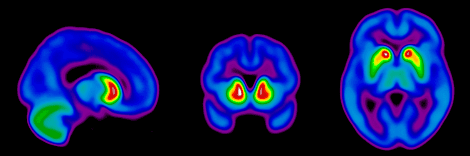
At baseline, DAT SPECT demonstrated significantly reduced striatal SBR in PD patients compared to healthy controls. VMAT2 PET data showed similar reductions, with improved spatial resolution and more specific signal localization. Longitudinal analysis revealed significant voxelwise reductions in striatal SBR for both DAT SPECT and VMAT2 PET.
Regional SBR analysis indicated that VMAT2 PET could detect significant changes as early as 12 months, comparable to DAT SPECT, despite lower data availability. Both imaging modalities showed the most significant changes in the putamen, with no distinct difference between the anterior and posterior putamen in the rate of decline.
Sample size estimates indicated that fewer participants were required to detect meaningful therapeutic effects using VMAT2 PET compared to DAT SPECT. For example, detecting a 60-80% reduction in striatal SBR change required fewer than 50 participants per arm with DAT SPECT at 24 months, whereas VMAT2 PET required fewer than 50 participants per arm as early as 12 months.
DAT SPECT and VMAT2 PET provide valuable insights into dopaminergic neurodegeneration in PD. While DAT SPECT is a robust and widely available diagnostic tool, VMAT2 PET offers superior spatial resolution and improved ability to detect disease progression. Both imaging biomarkers show promise in assessing disease-modifying therapies in PD, with VMAT2 PET demonstrating greater potential for the early detection of changes in dopaminergic function.
Presentation Highlights
This presentation has voice & text narration. You can mute/unmute the audio or open/close the transcript on this media player.