Quantitative Analysis of DaTscan SPECT Images without Anatomical MRI Reference
Reliable MRI-less DaTscan quantification to streamline Parkinson’s disease clinical trials.
Section 1
Key Takeaways
DaT SPECT is a well-established and widely available clinical tool that provides valuable insights into dopamine transporter density. Two different ways of processing the data to measure the SBR offers the possibility to use the processing technique better suited for the clinical study design.
Both DaT SPECT with subject-visit 3DT1 MRI registration and DaT SPECT without subject-visit 3DT1 MRI registration image processing can reliably track PD progression and may serve as valuable biomarkers in clinical trials evaluating disease-modifying therapies.
DaT SPECT with subject-visit 3DT1 registration and DaT SPECT without subject-visit 3DT1 registration image processing can be used in combination with clinical data for diagnostic confirmation during patient screening and randomization.
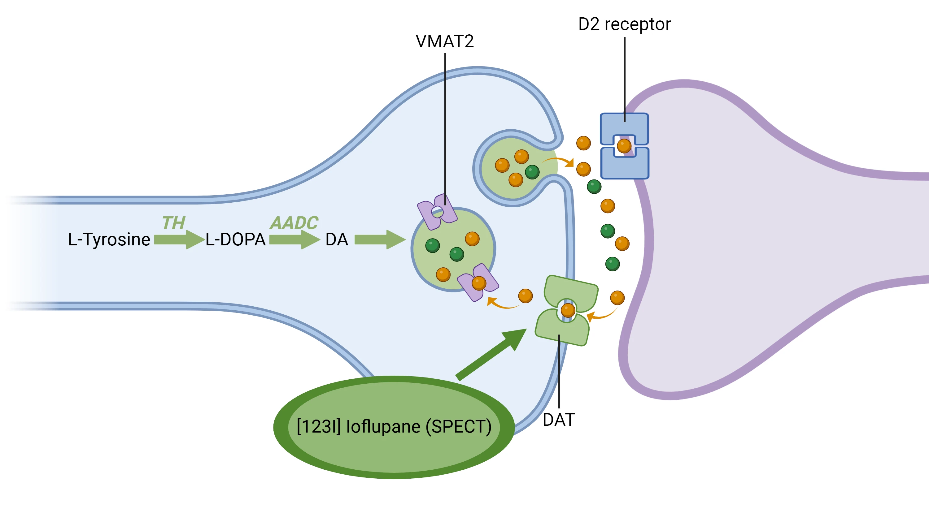
DaTscan ([123I]ioflupane) SPECT imaging enables visualization of presynaptic dopaminergic nerve terminals in the striatum by binding to the dopamine transporter (DaT). It is widely used to support the diagnosis of Parkinsonian syndromes, including Parkinson’s disease (PD), by differentiating them from conditions without dopaminergic deficits, such as essential tremor (ET). Quantitative analysis typically relies on the striatal binding ratio (SBR), defined as the ratio of specific to non-specific uptake, the latter typically based on the occipital lobe.
In clinical trials, SBR is frequently used for subject selection, stratification, randomization, and efficacy assessment. Accurate identification of dopaminergic deficit at screening is critical to minimize inclusion of phenocopies, common in early disease stages. Standard SBR estimation requires anatomical region-of-interest (ROI) definition, typically achieved by registering DaTscan images to subject-specific 3D T1-weighted (3DT1) MRI. However, MRI acquisition increases cost, logistical complexity, patient burden, and may be contraindicated or impractical in some participants. A DaTscan-only workflow could, therefore, improve accessibility and efficiency.
Our group previously developed a robust, fully automated DaTscan-to-MRI registration method based on rigid-body transformation, suitable for subject-specific anatomical alignment. In this work, we extend this approach to estimate a 9-parameter affine transformation, enabling registration of DaTscan images to a non-subject anatomical reference. Although template-based ROI placement may be affected by inter-subject anatomical variability, the relatively low spatial resolution of DaTscan SPECT images suggests that reliable SBR estimation remains feasible.
Using DaTscan SPECT and anatomical MRI data (305 scans from 101 subjects) from the Parkinson’s Progression Markers Initiative (PPMI) study, we compared SBR values derived with subject-specific MRI registration to those obtained using our “MRI-less”, template-based approach.
All scans processed successfully without subject MRI, relying solely on anatomical template built from 124 PPMI scans. The SBR values calculated with and without subject MRI showed excellent agreement, with a strong linear correlation (r = 0.94) using the occipital lobe as reference region.
MRI-less DaTscan SBR quantification using affine registration to an anatomical template is feasible and yields results comparable to MRI-based analysis. This approach will substantially reduce cost, logistics, and patient inconvenience, supporting broader adoption of DaTscan quantification in Parkinson’s disease clinical trials.
Slide Presentation
1/9
Related Content
Up-to-date information on best practices related to the use of neuroimaging in clinical trials of neurological diseases.
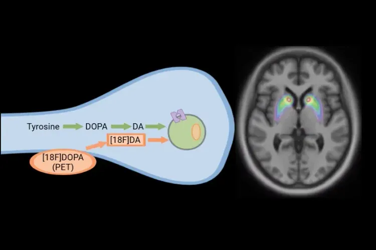
[18F]DOPA PET in Parkinson’s Disease Clinical Trials
How [18F]DOPA PET is used to monitor disease progression & response to therapeutic intervention in Parkinson’s disease & movement disorders clinical trials.
Computational Method for Harmonization of Brain PET Images
A new algorithm for standardizing the spatial resolution of brain positron emission tomography (PET) images in multicenter clinical trials without phantoms.
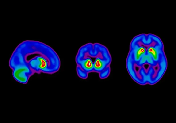
Dopaminergic Imaging in Parkinson's Disease
DAT SPECT and VMAT2 PET analysis in a natural history study of Parkinson's Disease.